Digital X-ray
Modern Diagnostics with Reduced Burden
Digital X-ray is an indispensable component of modern medical imaging today. It offers numerous advantages over conventional methods—both for diagnostics and for patient safety.
Procedure of the Examination
Depending on the body region to be examined, you will be asked to stand in front of the X-ray machine, sit, or lie down on the examination table. Our radiology technologists will ensure that you are positioned optimally—whether standing or lying down—to capture the best possible images of the area under examination. To protect your sensitive reproductive organs, you will be provided with lead shielding.
You may also receive instructions regarding your breathing or proper body positioning. You should remain still while the X-ray—which takes only a few seconds—is being taken. The X-rays pass through the body and are captured by a digital detector. Varying image contrasts are produced depending on tissue density.
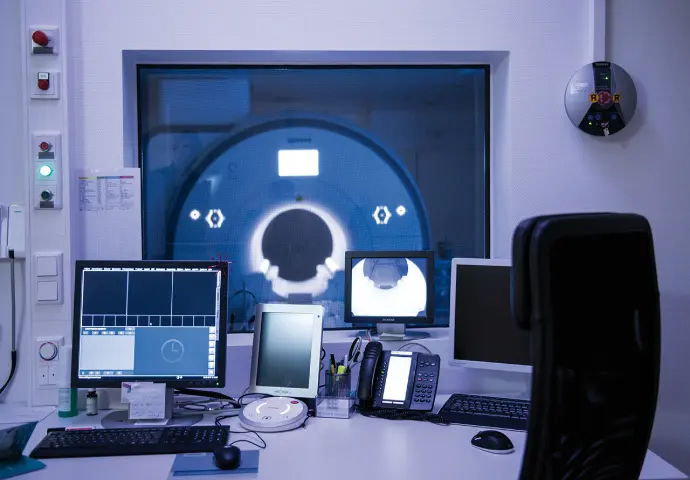
It is normal for the doctor and their assistants to leave the room during the examination to protect themselves from high radiation exposure. The X-ray examination takes only a few minutes in total.
Potential Risks
As with any X-ray examination, digital radiography also utilizes ionizing radiation. If used improperly or repeated frequently, this radiation can damage genetic material and slightly increase the risk of certain diseases, such as cancer.
The radiation dose during X-ray examinations is very low, which is why the risk is considered minimal. The X-ray examination itself is absolutely painless. Only the positioning—for example, of a broken foot—may be painful.
Radiation exposure in digital X-ray imaging is significantly lower than with conventional methods. Thanks to modern technology and optimized dose control, the exposure to the patient is reduced to a minimum.
State of the art
Digital radiography utilizes highly advanced detector systems that convert X-rays directly into digital image data. The advantages include a lower radiation dose, superior image quality, and faster availability.
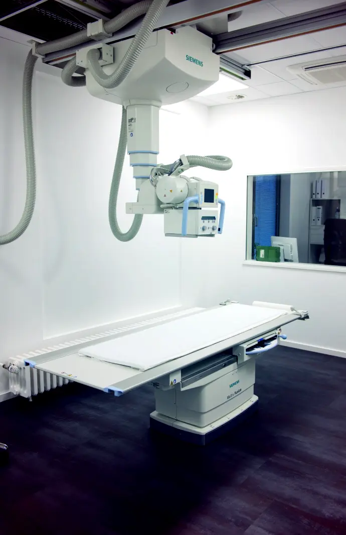
Our Siemens X-ray machines
With the Multix Fusion Digital Wireless, our practice "Radiology Düsseldorf Mitte" relies on a flexible and high-performance X-ray system that combines the highest image quality with intelligent dose reduction.
With the Multix Fusion Digital Wireless, our practice "Radiologe Viersen" relies on a flexible and high-performance X-ray system that combines the highest image quality with intelligent dose reduction.
Your Benefits at a Glance:
- Excellent image quality thanks to a high-resolution detector with cesium iodide technology.
- Intelligent dose reduction through copper filters, removable grids, and optimized table materials.
- Ergonomic design with a height-adjustable table, a free-floating tabletop, and extensive patient coverage (up to 190 cm).
- Wireless detector for maximum flexibility—even during examinations directly at the patient's bedside or in a wheelchair.
- Rapid image processing with DiamondView Plus for high-contrast and highly detailed visualization.
Siemens' digital X-ray systems align perfectly with our commitment to patient-centered care.
Patient-Information
Before the Examination
- Please inform us of a possible pregnancy, as X-rays can harm the unborn child.
- Wear comfortable clothing and, if possible, refrain from wearing jewelry, piercings, or metallic accessories in the examination area.
- Please bring any previous medical records or referral slips, if available.
- As a rule, medications do not need to be discontinued; however, please inform us of any relevant pre-existing conditions or allergies (e.g., to contrast agents).
During the Examination
- Follow the instructions of the medical staff, particularly regarding posture or breathing—this improves image quality.
- The examination is painless and usually takes only a few minutes.
- Remain calm and relaxed; unnecessary movements can impair recording quality.
After the Examination
- The report is prepared by the radiologist and sent to the referring physician.
- You may receive a CD containing your recordings, as well as access to the online patient portal.