Magnetic Resonance Imaging
Precise Imaging for Modern Diagnostics
Overview
Modern magnetic resonance imaging (MRI) uses a strong magnetic field to create precise cross-sectional images of the body.
This procedure is extremely effective for both diagnosing and ruling out diseases, and plays an important role in monitoring therapeutic progress. MRI is particularly important for examining the central nervous system, including the brain and spinal cord. Since it does not require the use of X-rays, it can be performed as often as necessary without posing any health risks to the patient.
Procedure of the Examination
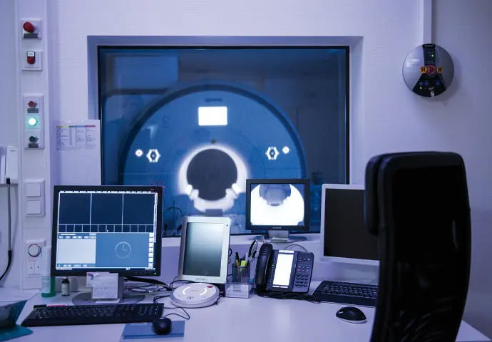
Before the examination, metallic objects must be removed. The patient lies on a table, which is slid into the MRI machine—a tube-shaped structure with built-in coils.
During the examination, you should remain as still as possible and breathe evenly, as movements can impair the quality of the images.
During the examination, loud knocking noises occur, which is why hearing protection is worn. The examination lasts between 10 and 25 minutes, depending on the body region.
Potential Risks
MRI is classified as very safe: there is no radiation exposure, such as that associated with X-ray or CT examinations. In rare cases, contrast agents can trigger allergic reactions.
Metallic objects in the body (e.g., implants, piercings) may heat up or impair image quality. Claustrophobia can be alleviated through the administration of sedatives.
State of the art
MRI technology is evolving rapidly. We offer optimized image processing utilizing the latest Siemens software, which enhances image quality and reduces examination times.
The optional AI-assisted reporting service powered by Floy® provides an additional analysis of your imaging data. The AI identifies potential risks or changes that are often difficult to detect in their early stages or require particularly complex analysis.
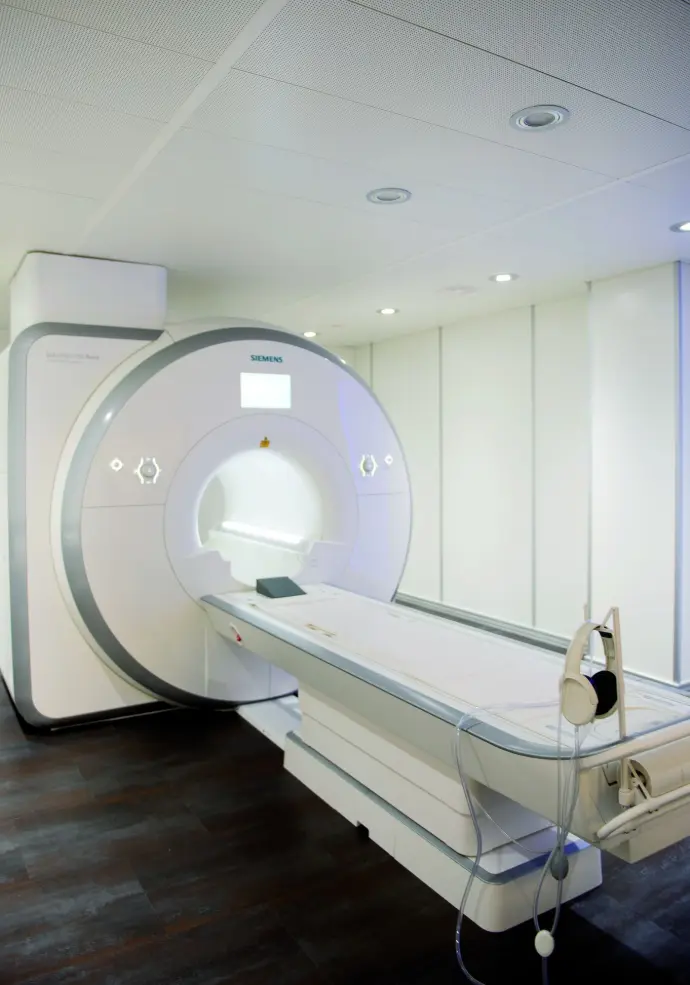
Our MRI: Siemens Magnetom Aera 1.5T
Our practice is committed to the highest standards of quality in diagnostic imaging—featuring the Siemens Magnetom Aera 1.5T, one of the most advanced MRI systems in its class. This system combines innovative technology with exceptional patient comfort, delivering razor-sharp images for precise diagnosis.
Special Benefits for Our Patients:
- Open design with a 70 cm gantry diameter: Ideal for people with claustrophobia, children, and patients with a higher body weight (up to 220 kg).
- Short examination times: Thanks to intelligent software and modern coil technology (Tim® 4G), many examinations are completed in just a few minutes.
- High image quality with maximum efficiency: The combination of powerful magnetic field strength (1.5 Tesla) and automated workflows ensures consistent and reliable results.
- Broad spectrum of applications: From neurological and orthopedic examinations to cardiac imaging and oncology—the Magnetom Aera covers virtually all clinical questions.
With the Magnetom Aera, we offer you modern, comfortable, and safe MRI diagnostics—tailored entirely to your needs.
Our MRI Services
We offer you a comprehensive spectrum of modern medical diagnostics and therapy—individualized, precise, and state-of-the-art.
Cardiac MRI (Heart Examination) / Angiography
With cardiac MRI and MR angiography, we offer radiation-free, high-precision imaging for the examination of the heart and blood vessels—enabling safe diagnosis and targeted treatment of heart diseases.
Small Intestine (Sellink)
Sellink MRI of the small intestine is a specialized, radiation-free examination used to visualize inflammation, strictures, or tumors in the small intestine—particularly helpful in cases of chronic inflammatory bowel diseases such as Crohn's disease.
Multiparametric Prostate MRI
Multiparametric prostate MRI is a modern, radiation-free procedure for the early detection of prostate cancer; it provides highly precise images and helps to identify suspicious tissue changes at an early stage and treat them in a targeted manner.
Mamma-MRI
Breast MRI is a radiation-free, highly sensitive examination of the breast capable of visualizing even the smallest changes—making it ideal for the early detection of breast cancer, particularly in cases of dense breast tissue.
MARS-MRI
MARS-MRI is a specialized MRI technique for the safe and low-artifact visualization of joints or soft tissues in patients with metal implants—entirely without radiation exposure.
Patient Information
Before the Examination
- Clothing: Wear comfortable, metal-free clothing. Wire-free bras and clothing without zippers or metal buttons are ideal.
- Remove metal items: Jewelry, watches, glasses, hearing aids, bank cards, etc., must be removed.
- Reporting Implants: You must inform the staff in advance if you have a pacemaker, cochlear implant, insulin pump, or metal fragments.
- Claustrophobia: Please let us know when scheduling your appointment. Sedatives may help.
During the examination
- Avoid movement, as it impairs image quality.
- Noise: Knocking sounds are normal—hearing protection is provided.
- Communication: Contact with staff is possible at any time via the intercom system.
- Contrast Medium: Used for specific diagnostic purposes (e.g., vascular diagnostics). Generally well tolerated; excreted via the kidneys. Please inform us in advance if you have any kidney conditions or allergies.
After the examination
- The report is prepared by the radiologist and sent to the referring physician.
- You may receive a CD containing your recordings, as well as access to the online patient portal.